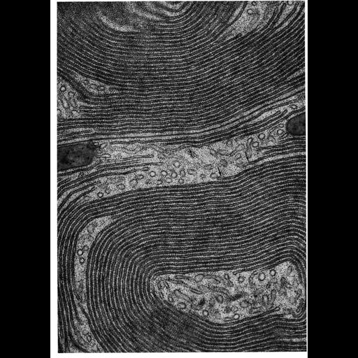
Figure 169 from Chapter 5 (Endoplasmic Reticulum) of 'The Cell, 2nd Ed.' by Don W. Fawcett M.D. Electron micrograph of rough endoplasmic reticulum (RER) in an acinar cell from the pancreas of the small brown bat, Myotis lucifugus. In this micrograph, the RER is composed of a concentric array of cisternal stacks, with successive layers about 35nm apart. Arrows indicate regions where the lumen widens, as is the case when the cisternae are continuous with tubular elements of the reticulum. This cisternal packed organization is common in cells that actively produce proteins for secretion. A PDF copy of the accompanying chapter is available on the ASCB’s BioEDUCATE website.
| Spatial Axis | Image Size | Pixel Size |
|---|---|---|
| X | 927px | —— |
| Y | 1289px | —— |