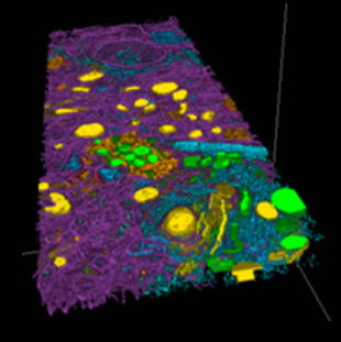
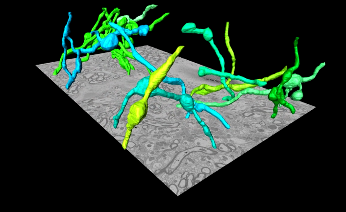
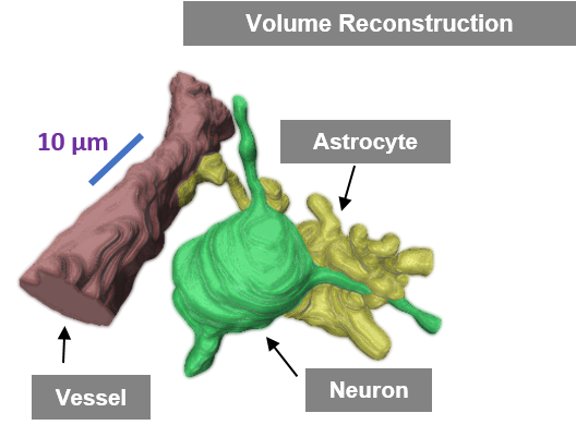
Reconstruction data
Final forms of cells and their structures are converted into mesh- or voxel-based renderings to visualize the structures in an image, as well as the high-resolution details of what subcecullar structures are inside of them as well as their tissue-context and what other cells they interact with.
These data-rich scenes display the attributes of the tissue, such as how cells appear in the tissue, how the subcellular contents of the cells organize, and how the pathological structures could be interfering. Studying the distribution of paired-helical filaments (PHF) and β-amyloid plaques within the sophisticated organization of neurons, blood vessels, and brain structures can lead to profound insights regarding the progression of AD.
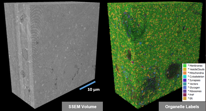
Models & Quantitative Data
Labeled image data is reduced into quantitative factors, and paired with trained A.I. models, presents the exact analysis workflow that was applied to yield a given set of quantative data that can help understand the underlying AD mechanisms.