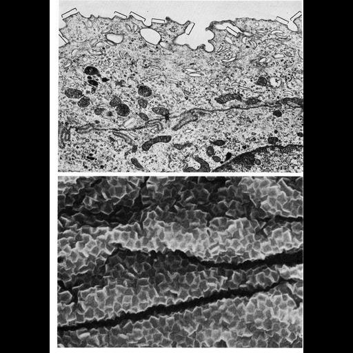
Transitional epithelial cells of the bladder. Upper panel is an electron micrograph from thin sectioned superficial cells of the uroepithelium in the rat; lower panel shows a scanning electron micrograph of the luminal surface of mouse bladder. Both images reveal rigid plaques (see brackets, top panel)along the surface of the epithelial tissue separated by more flexible interplaque regions. In the lower panel, the polygonal plaques appear sunken, while the more flexible interplaque regions appear as ridges or folds. Figures 11 (upper panel, courtesy of Marian Hicks) and 12 (lower panel, courtesy of Linda Malick) from Chapter 1 (The Cell Surface) of 'The Cell, 2nd Ed.' by Don W. Fawcett M.D. A PDF copy of the accompanying chapter is available on the ASCB's BioEDUCATE website.
| Spatial Axis | Image Size | Pixel Size |
|---|---|---|
| X | 914px | —— |
| Y | 1272px | —— |