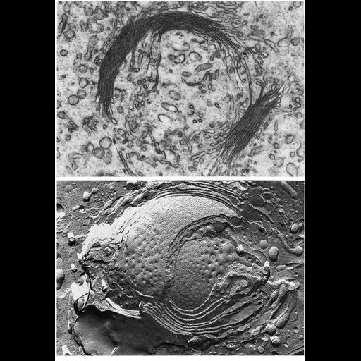
Figures 197 (upper) and 198 (lower) from Chapter 6 (Golgi Apparatus) of 'The Cell, 2nd Ed.' by Don W. Fawcett M.D. show electron microscopic views of the Golgi complex. Upper panel: A thin section preparation show the Golgi complex in the caudal cytoplasm of a ram spermatid. The cisternae are tightly packed, suggesting this organelle is relatively inactive. Lower: Freeze fracture replica of a more active Golgi complex from a guinea pig spermatocyte. Arrows point to expanded peripheral portions of the cisternae. A PDF copy of the accompanying chapter is available on the ASCB’s BioEDUCATE website.
| Spatial Axis | Image Size | Pixel Size |
|---|---|---|
| X | 900px | —— |
| Y | 1296px | —— |