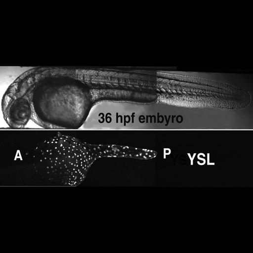
Visualization of yolk syncytial layer (YSL) nuclei in living zebrafish embryos. Sytox Green, a membrane-impermiant vital nuclear stain, was injected into the yold cell of late blastula (sphere stage) embryos. At 36 hrs of development, the zebrafish yolk cell displays a prominent anterior-posterior (A, P) axis, with brightly displayed fluorescent nuclei distributed through its entire cortex.
| Spatial Axis | Image Size | Pixel Size |
|---|---|---|
| X | 934px | —— |
| Y | 410px | —— |