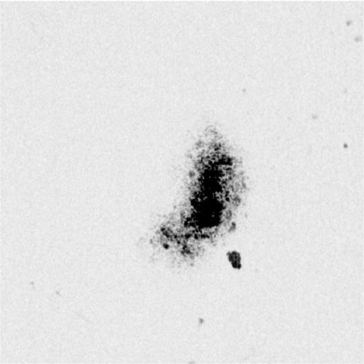
A wild-type mouse embryos on embryonic day 9.5 was isolated, fixed in 3.5% para-formaldehyde in PBS, rinsed in PBS, and was imaged with a prototype optical coherence microscopy (OCM) imaging system. With OCM an optical beam is directed at the tissue, and a small portion of this light that reflects from sub-surface features is collected. Note that most light is not reflected but, rather, scatters. Scattered light can be filtered out using optical coherence. Only the reflected (non-scattered) light is coherent (i.e., retains the optical phase that causes light rays to propagate in one or another direction). In the OCM, an optical interferometer is used to detect only coherent light. The OCM data was acquired over a 400um x 400um field of view at 2 frames per second. The spacing between each 3D-OCM slice is 5um along the depth dimension. The OCM data on the mouse embryo confirmed that myocardial-endocardial radial associations exist in the intact ED 9.5 looping mouse heart.
| Spatial Axis | Image Size | Pixel Size |
|---|---|---|
| X | 700px | 0.57µm |
| Y | 700px | 0.57µm |
| Z | 102px | 5µm |