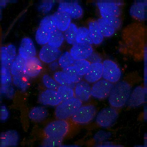
FISH detection of ERBB2 (florescein, green) and MYC (rhodamine, red) in normal breast tissue. Nuclei counterstained with DAPI (blue). Tissue was fixed in formaldehyde, embedded in paraffin, and 4-5 micron sections cut. Stained sections were viewed using an Olympus IX70 microscope with a 60x 1.4 NA objective lens, and images acquired with a Photometrics CoolSnap CCD with a 1024X1024 pixel resolution, with a pixel size equivalent to 0.07427 microns in X and Y. Shown is a maximum intensity projection of the stack. The complete stack is included in this group of images Part of this field of view is shown in Fig 1B of Meaburn et al., 2009, J Cell Biol 187:801-812.
| Spatial Axis | Image Size | Pixel Size |
|---|---|---|
| X | 1024px | 0.074µm |
| Y | 1024px | 0.074µm |