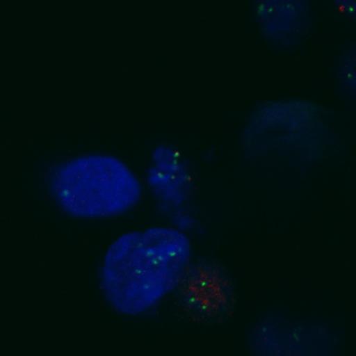
This image is part of Fig. 2B from the reference article PMID 20212317. HeLa cells were treated with control siRNA and double thymidine block for 48 h. Cells were fixed with 4% paraformaldehyde, dehydrated 100% methanol at −20°C, and stained for centromeres (CREST, red, Alexa Fluor 568 conjugated secondary Ab), Bub1 (green, Alexa Fluor 488 conjugated secondary Ab) and Hoechest 33342 (blue). Fluorescence microscopy was performed at RT on a confocal microscope (LSM510 Meta; Carl Zeiss, Inc.) equipped with a 100× Plan-Apochromat objective. A 543 nm HeNe laser (5 mW output; detection LP560 nm) was used for detection of Alexa Fluor 568–labeled antibodies. The 488nm line of an Argon laser (25 mW nominal output; detection BP 505–530 nm) was used for analysis of Alexa Fluor 488–labeled antibodies. Hoechst 33258 images were captured using the 364nm line of an ion laser (Enterprise II ML UV; Coherent, Inc.; 80 mW nominal output; detection BP 385–470 nm).
| Spatial Axis | Image Size | Pixel Size |
|---|---|---|
| X | 1024px | —— |
| Y | 1024px | —— |
| Z | 19px | —— |