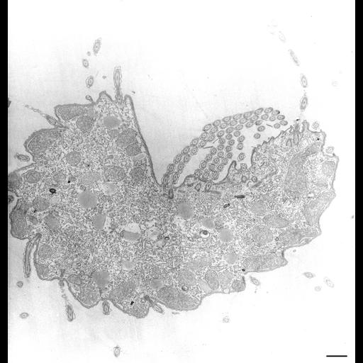
A cross section of the cell (ventral side up) through the buccal cavity showing basal bodies of the 3 membranelles (1, 2 and 3) lying along the indented dorsal wall. A short segment of the oral ribs separates the membranelles from the undulating membrane that lies at the margin of the right wall. Basal body/cilium complexes of a kinety are found at the margin of the left wall. The left wall is covered by ER on its cytosolic side. TEM taken on 8/25/67 by R. Allen with Philips 200 operating at 60kV. Neg. 4,500X. Bar = 1µm. The negative was printed to paper and the image was scanned to Photoshop. This digitized image is available for qualitative analysis. There is a high resolution version of this image in the library (CIL:39209) which is available for quantitative analysis. Additional information available at (http://www5.pbrc.hawaii.edu/allen/).
Standard glutaraldehyde fixation followed by osmium tetroxide, dehydrated in alcohol and embedded in an epoxy resin. Microtome sections prepared at approximately 75nm thickness. Additional information available at (http://www5.pbrc.hawaii.edu/allen/).
| Spatial Axis | Image Size | Pixel Size |
|---|---|---|
| X | 3197px | —— |
| Y | 3336px | —— |