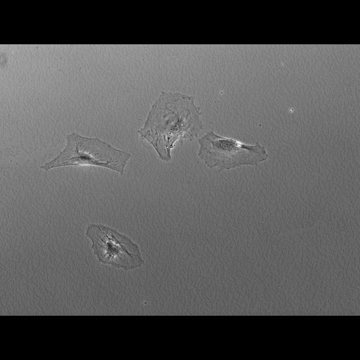
Time lapse movie of mouse embryonic fibroblasts in culture imaged at 30 second intervals by phase contrast microscopy. A micropipette is positioned near the cells to deliver either a growth factor or control buffer. In this sequence when a growth factor is added the cells activate a ring of ruffles that begin at the periphery and converge most likely at the site of the primary cilium followed by whole cell motility towards the pipette tip.
These data were collected as research which led to the published article Cell Physiol Biochem 2010;25:279-292. Microscopy was performed with a 20X planapo phase contrast objective on an Olympus IX 70 microscope with a SensicamHQ cooled CCD camera and a heated stage insert set to 37 degrees C. Cells were grown and serum starved for 48 h in a coverslip bottom MatTek dish. Small amounts of PDGF-BB were continuously ejected from the micropipette to create a gradient in the vicinity of growth-arrested wt MEFs. Cell movement was monitored with time lapse video microscopy, taking images at 30 second intervals. A Femtojet Micromanipulator 5171 (Eppendorf-Brinkman Instruments) and a pump (model Femtojet; Eppendorf) were used to control the position of the micropipette and the pressure required for the chemoattractant flow. Femptotip II micropipettes were positioned within 1 μm of the cover slip and pressure set at 30 to 45 hPa. Spatial scale is approximate.
| Spatial Axis | Image Size | Pixel Size |
|---|---|---|
| X | 1280px | 0.33µm |
| Y | 1024px | 0.33µm |
| Time | 30 seconds | 512 |
|---|