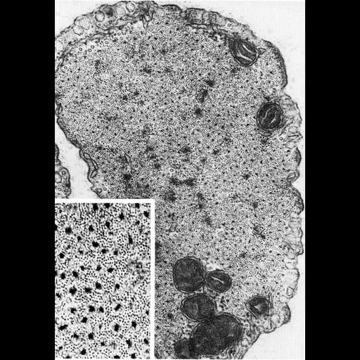
Figure 434 from Chapter 16 (Cytoplasmic matrix and cytoskeleton) of 'The Cell, 2nd Ed.' by Don W. Fawcett M.D. Vascular smooth muscle cell in cross section shows myosin and actin filaments distributed throughout the cytoplasm. In the higher magnification inset panel, thick myosin filaments are surround by thinner actin filaments, outnumbering the myosin filaments by about 15:1. Image from Andrew Somlyo, Vascular Smooth Muscle, Springer-Verlag, Heidelberg, 1972. A PDF copy of the accompanying chapter is available on the ASCB’s BioEDUCATE website.
| Spatial Axis | Image Size | Pixel Size |
|---|---|---|
| X | 897px | —— |
| Y | 1290px | —— |