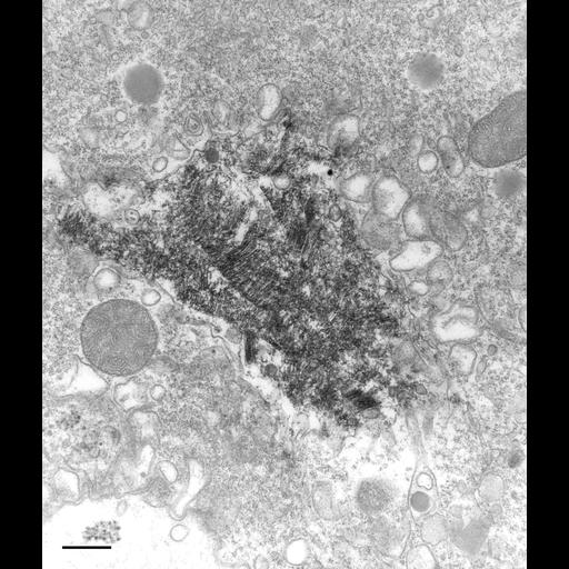
This micrograph shows a crystal surrounded with secondary lysosomes. This was taken from a cell returned to growth medium following starvation for several days (0 day of rejuvenation). This could represent either fusion of lysosomes with a growing crystal or reformation of secondary lysosomes from a crystal when the cell has been returned to a culture with an abundant food supply. TEM taken on 2/12/81 by R. Allen with Hitachi HU11A operating at 75kV. Neg. 12,750X. Bar = 0.5µm.
Standard glutaraldehyde fixation followed by osmium tetroxide, dehydrated in alcohol and embedded in an epoxy resin. Microtome sections prepared at approximately 75nm thickness. The negative was printed to paper and the image was scanned to Photoshop. This digitized image is available for qualitative analysis. Additional information available at (http://www5.pbrc.hawaii.edu/allen/).
| Spatial Axis | Image Size | Pixel Size |
|---|---|---|
| X | 2557px | —— |
| Y | 3000px | —— |