Alternate header for print version


Licensing
Public Domain: This image is in the public domain and thus free of any copyright restrictions. However, as is the norm in scientific publishing and as a matter of courtesy, any user should credit the content provider for any public or private use of this image whenever possible. Learn more
tweet  
*CIL – Cell Image Library accession number. Please use this to reference an image.

CIL:36723*  Cite 
Description

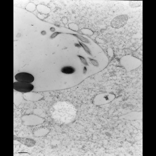
Monoclonal antibody, E30-2-1, was raised to antigen D6 that specifically labels acidosomes and DV-II. There was no label on the nascent vacuole membrane or ciliary membranes. This cell had been incubated in 1.1µm latex beads for 3 minutes and fixed without a chase. There is some gold over the cytosol but the nascent vacuole lumen is essentially free of label. TEM taken on 7/12/90 by R. Allen with Zeiss 10A operating at 80kV. Neg. 9,780X. Bar = 0.5µm.

Technical Details

To label membranes inside the cell we used very lightly fixed cells (0.25% glutaraldehyde) that were then rapidly frozen in liquid nitrogen and sectioned later at -100oC. These frozen sections were picked up on drops of methylcellulose and transferred to a Formvar-supported grid. The sections were immunogold labeled (15nm gold) to show the location of the specific antigen inside the cell as well as on the cell surface. Microtome sections prepared at approximately 75nm thickness. The negative was printed to paper and the image was scanned to Photoshop. This digitized image is available for qualitative analysis. Additional information available at (http://www5.pbrc.hawaii.edu/allen/).

Biological Sources
NCBI Organism Classification
Paramecium multimicronucleatum
Cell Type
cell by organism
eukaryotic cell
Eukaryotic Protist
Ciliated Protist
Cellular Component
food vacuole
early phagosome
Biological Context
Biological Process
digestive system process
Attribution
Names
Richard Allen (University of Hawaii)
Citation
Digital Object Identifier (DOI)
doi:10.7295/W9CIL36723
Archival Resource Key (ARK)
ark:/b7295/w9cil36723
Imaging
Image Type
transmission electron microscopy (TEM)
illumination by electrons
Image Mode
detection of electrons
film
Parameters Imaged
electron density
intrinsic mass distribution
Source of Contrast
stain with broad specificity
Visualization Methods
stain with broad specificity
uranyl salt
gold
Processing History
recorded image
film
Print from negative scanned for Photoshop.
Data Qualifiers
processed data
Dimensions
Spatial Axis Image Size Pixel Size
X 2268px ——
Y 2736px ——