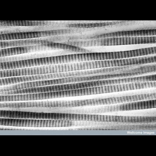
Transmission electron micrograph showing collagen fibrils in longitudinal view from a patient with glaucoma. The a-e banding is visualized by uranyl acetate staining of charged amino acid residues in the constituent collagen molecules. This specimen is also stained with a cationic dye to reveal interfibrillar proteoglycans. Although the fibrils look normal these proteoglycans are reduced in this area supporting the theory that abnormal proteoglycans are a cause of glaucoma.
B0001808 Collagen fibrils. Wellcome Images available under the following creative commons usage http://creativecommons.org/licenses/by-nc-nd/2.0/uk/
| Spatial Axis | Image Size | Pixel Size |
|---|---|---|
| X | 782px | —— |
| Y | 576px | —— |