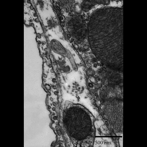
Transmission electron micrograph of coated vesicles in mouse cardiac tissue. Coated vesicles have the materials bound to the receptor molecules incorporated within their membranes allowing the vesicle to pick up and drop off material continuously re-use, this is called receptor-mediated endocytosis.
Primary fixation included: 2.5 % glutaraldehyde, 2% formaldehyde in 0.1 M Na-phosphate buffer, pH 7.4. Post-fixed in 2% OSO4 in 0.1 M Na-phosphate buffer, pH 7.4. Stained en bloc in 1% uranyl acetate. The tissue was then dehydrated in a graded series of ethanol and infiltrated with Spurr’s resin. Thin sections of 70 nm were trimmed using a diamond knife and post-stained in uranyl acetate and lead citrate. This micrograph image was taken using a Phillips CM 100 transmission electron microscope at an accelerating voltage of 80kV.
| Spatial Axis | Image Size | Pixel Size |
|---|---|---|
| X | 1252px | —— |
| Y | 1850px | —— |