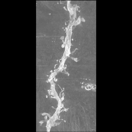
Maximum intensity projection of a tomographic reconstruction of a selectively stained spiny dendrite from a striatal medium spiny neuron from the neostriatum of a wildtype mouse. This reconstructed image has been downsampled from the raw data image, which can be accessed using the link provided to the Cell Centered Database.
Individual cells in fixed tissue were injected with Lucifer yellow and stored in 4% paraformaldehyde at 4° C. Fluorescence was photoconverted using DAB and a confocal laser light source. After this process, tissue was washed thoroughly in PBS and processed for EM by osmication, dehydration, and embedding in epoxy resin. For detailed methods, see the protocol provided in the CCDB. The tomogram was generated using an Hitachi UHVEM. Single tilt images spanned -66 to 66° in 2° increments.
| Spatial Axis | Image Size | Pixel Size |
|---|---|---|
| X | 512px | —— |
| Y | 512px | —— |