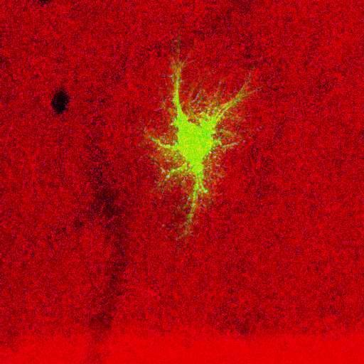
Projection through merged optical section series of a filled astrocyte (green) in the molecular layer of the dentate gyrus of a 1 month-old rat, stained for NCAM (red), showing the relationship between astrocyte processes and laminar boundaries revealed by NCAM staining. Optical sections were generated with a single photon confocal microscope. Original data contributed to Bushong EA, Martone ME, Ellisman MH. Examination of the relationship between astrocyte morphology and laminar boundaries in the molecular layer of adult dentate gyrus. J Comp Neurol. 2003 Jul 21;462(2):241-51. PMID: 12794746. This image has been downsampled from the raw data image which can be accessed using the link provided to the Cell Centered Database.
Projection through merged optical section series of a filled astrocyte (green) in the molecular layer of the dentate gyrus of a 1 month-old rat, stained for NCAM (red), showing the relationship between astrocyte processes and laminar boundaries revealed by NCAM staining. Optical sections were generated with a single photon confocal microscope. Original data contributed to Bushong EA, Martone ME, Ellisman MH. Examination of the relationship between astrocyte morphology and laminar boundaries in the molecular layer of adult dentate gyrus. J Comp Neurol. 2003 Jul 21;462(2):241-51. PMID: 12794746. This image has been downsampled from the raw data image which can be accessed using the link provided to the Cell Centered Database.
| Spatial Axis | Image Size | Pixel Size |
|---|---|---|
| X | 512px | —— |
| Y | 512px | —— |