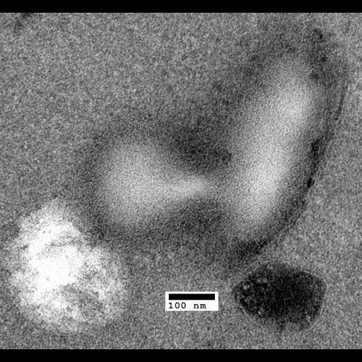
Transmission electron micrograph of a pair of lipid droplets in the act of fusion or fission. This image was taken in a sertraline-treated swa2 mutant yeast cell. (25,000X). CIL 40426 shows more detail of the membrane encapsulating the lipid droplet in similarly treated mutant cells.
This group of images contains representative whole-cell images and high-magnification zooms of organelle structures of interest. All cells were grown in liquid rich media (YPD pH6.5) at 30C with aerated shaking, and cultures were sampled in mid-logarithmic phase. Treated cells were exposed to 60µM antidepressant sertraline (Zoloft®) for 45 minutes.
| Spatial Axis | Image Size | Pixel Size |
|---|---|---|
| X | 1804px | —— |
| Y | 1680px | —— |