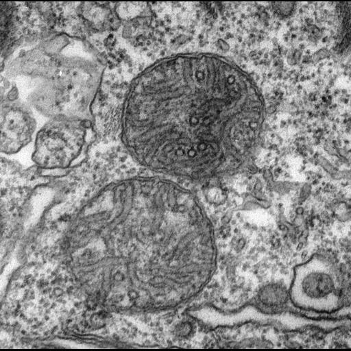
Enlargement of two mitochondria showing the cross sections of tubular cristae bear one pair of protrusions (these are the F1 subunits of F1F0 ATP synthase enzymes) while rows of protrusions can be viewed along some tangentially sectioned tubules. The tubules in some cases can be seen to arise from the inner membrane. TEM taken on 3/7/80 by R. Allen with Hitachi HU11A operating at 75kV. Neg. 22,500X. The raw negative was scanned with an Epson Perfection V750 Pro and this high resolution image is best used for quantitative analysis. Additional information available at (http://www5.pbrc.hawaii.edu/allen/).
Standard glutaraldehyde fixation followed by osmium tetroxide, dehydrated in alcohol and embedded in an epoxy resin. Microtome sections prepared at approximately 75nm thickness. The negative was printed to paper and the image was scanned to Photoshop. This digitized image is available for qualitative analysis. Additional information available at (http://www5.pbrc.hawaii.edu/allen/).
| Spatial Axis | Image Size | Pixel Size |
|---|---|---|
| X | 1819px | 0.89nm |
| Y | 1803px | 0.89nm |