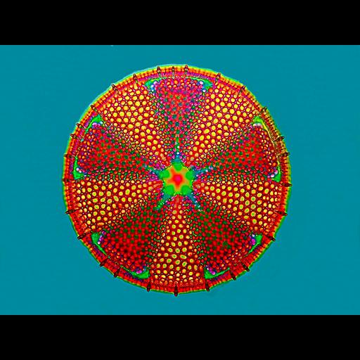
Fossil of an extinct diatom from Tertiary deposits in Dunkirk, Maryland. The diatom is approximately 80 microns in diameter. The image is of a diatom valve (two valves and a girdle band compose a diatom frustule or shell). The image was captured using Zeiss Jamin-Lebedeff Interference Contrast with a 40x objective. The image is of a diatom valve. Digital image stacking was also used to provide a greater depth of focus than would be possible with a single exposure. It is a stack of three separate exposures processed through Helicon Focus, which extracts pixels in focus in each image and then outputs a composite. Third Prize, 2008 Olympus BioScapes Digital Imaging Competition®.

| Spatial Axis | Image Size | Pixel Size |
|---|---|---|
| X | 3072px | —— |
| Y | 2304px | —— |