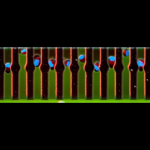
This confocal and phase contrast image depicts the chemotactic behaviour of MDA-MB-231 breast cancer cells. The nuclei are labelled blue and the mitochondria are labelled red. The cells are squeezed into micro-scale channels to study large numbers of single cells migrating with varying concentrations of epidermal growth factor (EGF, shown in green) at the leading and trailing edge of the cell. The individual channels are 12 microns wide. Wellcome Image Award 2012.
0008497. 2011 Collection: Wellcome Images Copyrighted work available under Creative Commons by-nc-nd 2.0 UK: England & Wales, see http://images.wellcome.ac.uk/indexplus/page/Prices.html
| Spatial Axis | Image Size | Pixel Size |
|---|---|---|
| X | 1476px | —— |
| Y | 550px | —— |