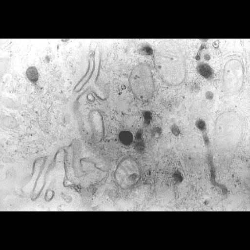
Human duodenal epithelium, peroxisomes contrasted with DAB by their catalase activity at pH 10.5, postosmicated, embedded in epon resin, and visualized using electron microscopy. Image shows two types of peroxisomes: large rounded structures, and elongated ones with a smaller diameter. More details in Roels et al: http://gut.bmj.com/content/32/8/858.long