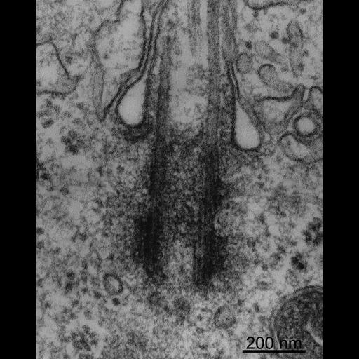
The electron micrograph shows the electron dense basal body in a testis cell in primary culture.
Testes from 15-day-old Sprague-Dawley rats (Sasco, Omaha Neb.) were dissociated in 0.1% trypsin and plated in NCTC-135 in Petri dishes containing cover slips. Cells on cover slips were fixed in 2.5% glutaraldehyde, 1% osmium tetroxide, stained en bloc with uranyl acetate, dehydrated and embedded in Araldite. Sections were taken parallel to the cover slip, stained with uranyl acetate and lead citrate and examined with a Philips 300 transmission electron microscope. Images were captured on film, printed onto photographic paper, digitized with a scanner and processed with Photoshop (Adobe).
| Spatial Axis | Image Size | Pixel Size |
|---|---|---|
| X | 4546px | —— |
| Y | 5671px | —— |