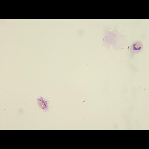
A monocyte contained a rounded zone resembling to aglutinated erythrocytes (upper half of the photo). Three partially aglutinated erythrocytes were at contact of a lytic nuclear-type material. The cytoplasm was almost not visible.
Cerebrospinal fluid, cytocentrifugation, May-Grunwald-Giemsa stain.