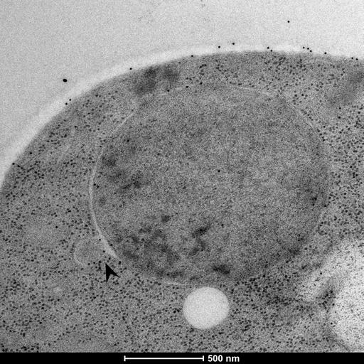
Heat shocked Saccharomyces cerevisiae cells (38 oC for up to 90 minutes) were high pressure frozen and embedded in HM20 resin. Samples were then stained for Ubiquitin with the polyclonal antibody ab19247 (Abcam) and visualized through transmission electron microscopy (TEM). The images show the nucleus of the cells and the black arrow indicates nuclear envelope budding events. The black gold particles represent the presence of Ubiquitin.
Heat shocked cells were cultured in YPD medium and high pressure frozen in a Wohlwend Compact 3 machine, followed by a short freeze substitution protocol in 2% Uranyl acetate and embedded in HM20 resin. For the immuno-EM, samples were blocked for 2 hours in 0.8% BSA with 0.1% fish skin gelatine. A 1:10 dilution of the primary antibody ab19247 (Abcam) was used and a 1:20 dilution of the secondary antibody anti-Rabbit IgG 10 nm gold (Cat#25108, Electron microscopy sciences). Finally, samples were post fixed in 2.5% glutaraldehyde for 20 minutes. Sections of 70 nm thickness were then contrast stained with 2% Uranyl acetate and Reynold's lead citrate. Pictures were aquired in a Tecnai T12 TEM with a Ceta CMOS 16M camera.