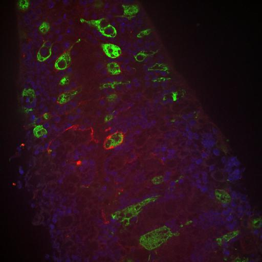
Confocal image of the CNS of a D. melanogaster in the third instar larval stage after αSNAP KD, 8uM from the ventral surface, depicting astrocytes (red), cortex glia (green) and neuronal nuclei (blue). Our study examined the breakdown of the spatial segregation between cortex glia and astrocytes after disruption of cortex glial morphology. After knockdown of αSNAP, a protein part of the vesicual transport pathway, we observed aberrant infiltration by astrocytes into the cortex.
Astrocyte-specific RFP expression was achieved using driven using the Gal4-UAS binary system and the driver alrm. Cortex-glia specific GFP was achieved using the lexA-lexAop binary system and the cortex-glia specific driver wrapper932i-LexA (Coutinho-Budd et al., 2017). The larval CNS was dissected in the third instar larval stage, fixed with methanol, stained for RFP, GFP and Elav (neuronal nuclei marker), and mounted in VectaShield reagent. Slides were imaged on an Innovative Imaging Innovations (3i) spinning disk confocal microscope equipped with a Yokogawa CSX-W1 using a 63X oil objective.