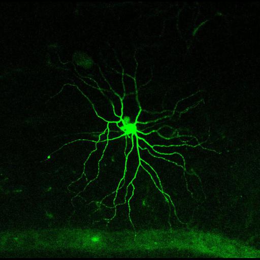
The photopigment melanopsin confers photosensitivity upon a minority of retinal output neurons. These intrinsically photosensitive retinal ganglion cells (ipRGCs) are more diverse than once believed, comprising five morphologically distinct types, M1 through M5. Imaged here is an M2 subtype as described in Estevez et al., J Neurosci. 2012
These color images show the three-dimensional morphology of an M2 melanopsin retinal ganglion cell from an ex-vivo isolated flat-mount mouse retina preparation. This retinal neuron was visually identified and targeted for dye injection based on somatic EGFP fluorescence expression the Opn4cre/+;Z/EG+/- mouse line as previously described (Ecker et al., 2010, Neuron). Lucifer yellow dye (green) was injected into the cell to reveal dendritic morphology of the M2 cell.
Images were acquired with a Zeiss LSM 510 Meta laser scanning microscope with a plan-apochromat 20x/0.8 objective.
The confocal image stack consisted of a 450 µm x 450 µm window through 14 stacks at 0.5 µm (total of 8.5 µm in z-depth) through the ganglion cell layer and ON-sublayer of the inner plexiform layer of the retina.
| Spatial Axis | Image Size | Pixel Size |
|---|---|---|
| X | 1024px | 450µm |
| Y | 1024px | 450µm |
| Z | 17px | 8500nm |